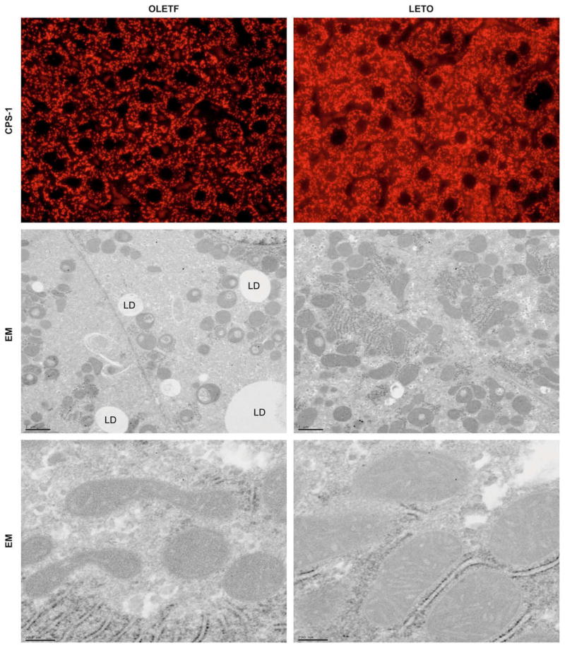
Fig. 5. Representative immunofluorescent photomicrographs from liver for mitochondrial marker CPS-1 (red) shows granular staining patterns in liver sections (A).
While there were no differences between animal groups at 5, 8, 13, and 20 weeks of age (not shown), the OLETF livers (left) had visibly decreased CPS-1 staining compared to the LETO animals (right) at 40 weeks of age. Also shown are representative electron micrographs (EM) from liver of OLETF (left) and LETO (right) rats at low (B) and high (C) magnifications. While no apparent differences were observed at 5, 8, or 13 weeks of age (data not shown), the mitochondria in the OLETF animals at 20 and 40 weeks show signs of rounding (B) and disruptions in cristae and outer and inner mitochondrial membranes (C). LD = lipid droplet.

