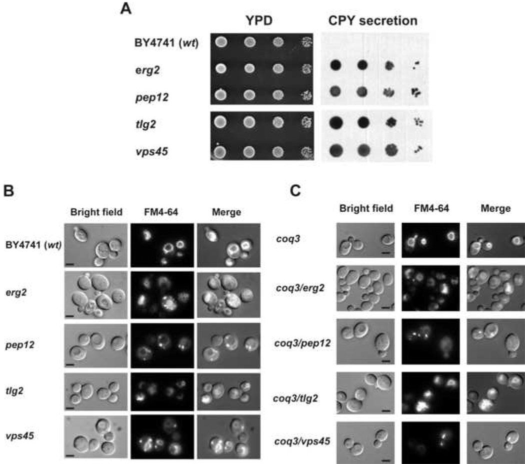
Figure 4.
Analysis of traffic membrane markers. Panel A: CPY secretion in single membrane traffic mutants. Equal number of cells from freshly grown yeast cultures were deposited as undiluted, 1:10, 1:100 or 1:1000 dilutions (left to right) on a nitrocellulose filter overlaid on YPD plates. The plates were incubated for 24 hours at 30 °C and washed as described under Experimental Procedures. Extracellular CPY secreted from colonies was detected by inmunostaining with anti-CPY antibody (Molecular Probes). Panel B: FM4-64 vacuolar staining. Yeast cell grown to logarithmic phase in YPD were incubated with 2 µM FM4-64 at 30°C during 30 min. After incubation, cells were washed with PBS and observed under the fluorescence microscope. Results are representative of a set of three experiments. Micrographs are obtained with 1000x magnification. Bar, 5 µm. Panel C: FM4-64 vacuolar staining in membrane traffic/coenzyme Q biosynthesis double mutants. Yeast cell grown to logarithmic phase in YPD were incubated with 2 µM FM4-64 at 30°C during 30 min. After incubation, cells were washed with PBS and observed under the fluorescence microscope. Results are representative of a set of three experiments. Micrographs are obtained with 1000× magnification. Bar, 5 µm.

