Fig. 3.

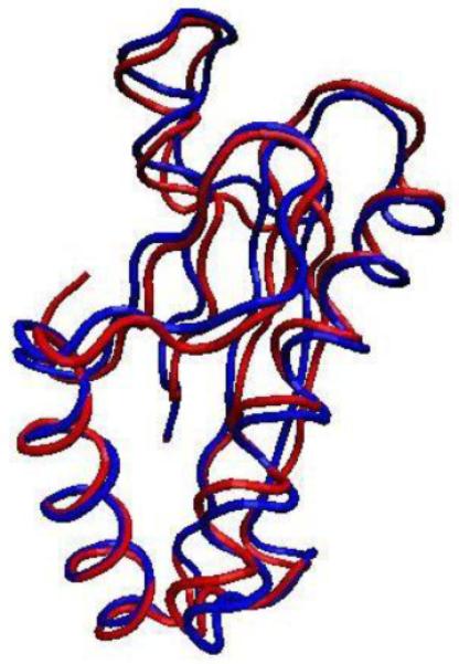
Backbone representations of truncated P6 from NTHi (blue, PDB ID 2AIZ) and Pal from E. coli (red, PDB ID 1OAP) overlay with an RMSD of ~1.2 Å. The graphical representation was prepared using the Visual Molecular Dynamics program [31].

Backbone representations of truncated P6 from NTHi (blue, PDB ID 2AIZ) and Pal from E. coli (red, PDB ID 1OAP) overlay with an RMSD of ~1.2 Å. The graphical representation was prepared using the Visual Molecular Dynamics program [31].