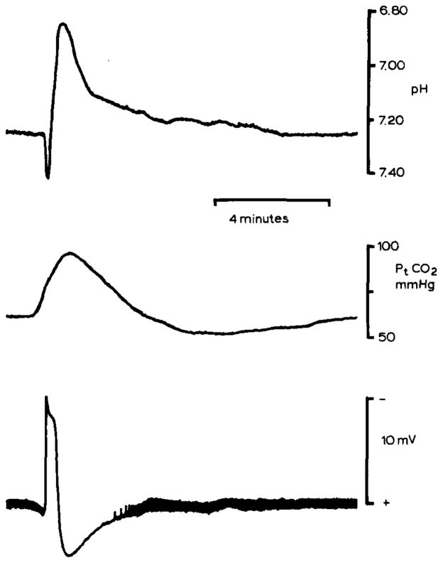
Fig. 3.
pHo and Pt(CO2) changes during spreading depression in rat neocortex. pHo (upper record) was recorded 500–600 μm below the pial surface in parietal neocortex with a double-barrelled H+ selective microelectrode. pHo shifted initially alkaline and then acid as in the cerebellum. However, the peak acid shift reached 6.90 pH which may reflect enhanced lactic acidosis since blood glucose had been elevated to 17 mmol/l by previous intraperitoneal injection of dextrose. Spreading depression occurred spontaneously. Middle record shows simultaneous recording of Pt(CO2) from a surface carbon dioxide microelectrode. Since the time constant of the carbon dioxide microelectrode is greater than about 30 seconds, peak Pt(CO2) may be artifactually low. Nonetheless, these Pt(CO2) changes show that brain activity can crease local inhomogeneities in tissue CO2 tension as well as [H+]o. Lower record shows the slow d.c. potential negative shift associated with spreading depression. Note that the thickness of the baseline reflects spontaneous electrical activity of the cortex which stops during spreading depression and then progressively returns. These data were recorded during experiments described in Kraig et al., 1984b, 1985c.

