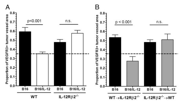
FIGURE 3.
IL-12 does not act directly on endothelial cells or tumor cells to mediate VEGFR3 suppression on tumor vessels. A, B16 or B16/IL-12 cells (2 × 10⁵) were injected i.m. into WT or IL-12Rβ2−/− mice and grown to a mean thigh diameter of 10–13 mm. B, Lethally irradiated WT or IL-12Rβ2−/− mice were reconstituted with WT or IL-12Rβ2−/− bone marrow, rested for 8 wk, and injected i.m. with 2 × 10⁵ B16 or B16/IL-12 cells. Tumors were then removed as stated in the Materials and Methods, stained for VEGFR3 and CD31, and visualized by whole mount fluorescence microscopy. The proportion of vessel area that stained positive for VEGFR3 was calculated using ImagePro software. The dotted line at 0.36 represents the level of staining seen in B16/IL-12 tumors grown in a WT mouse, which does not have VEGFR3 on its vessels and therefore represents nonspecific staining. Graphs represent two experiments with four to six animals per condition. Significance was calculated using Bonferroni’s multiple comparison test.

