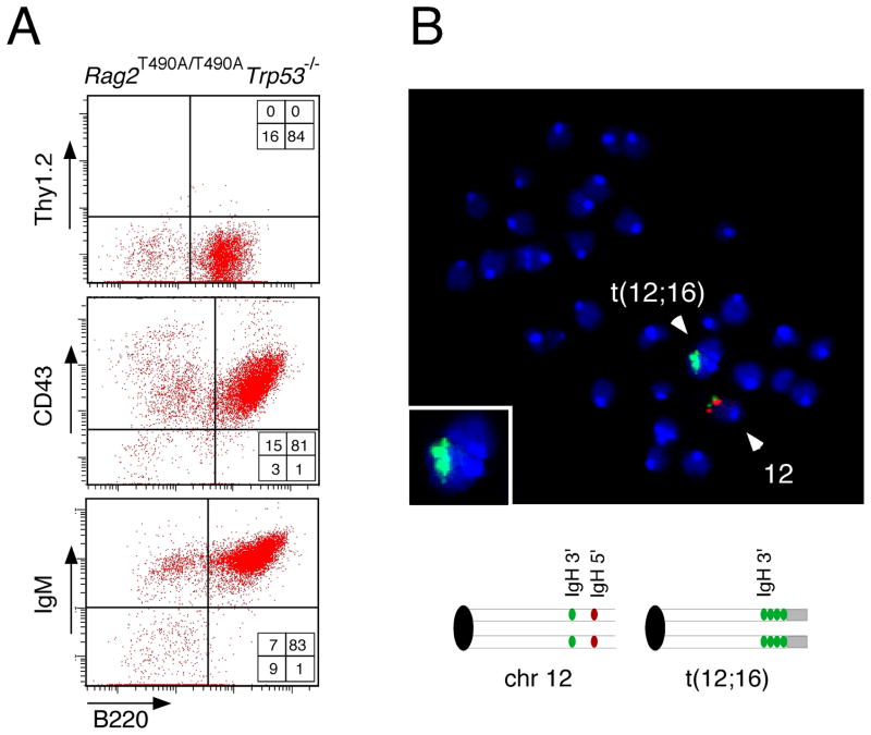
Figure 6.
A t(12;16) translocation splits and amplifies a portion of the IgH locus in a B cell lymphoma from a p53-deficient, RAG-2 T490A mouse. (A) Flow cytometric analysis of tumor 2135 (Rag2T490A/T490ATrp53−/−) indicates a tumor of B lymphoid origin. Dispersed tumor cells were stained for the surface markers B220 and Thy1.2 (top), CD43 (middle) and IgM (bottom). (B) Analysis of a metaphase spread from the same tumor by FISH using BAC probes specific for the 5′ flank of the IgH locus (red) and the 3′ flank of the IgH locus (green). Inset represents an enlarged image of t(12;16), which had been detected by SKY analysis (see Table 1). The deduced arrangements of probes in intact chromosome12 and the t(12;16) translocation are indicated below.

