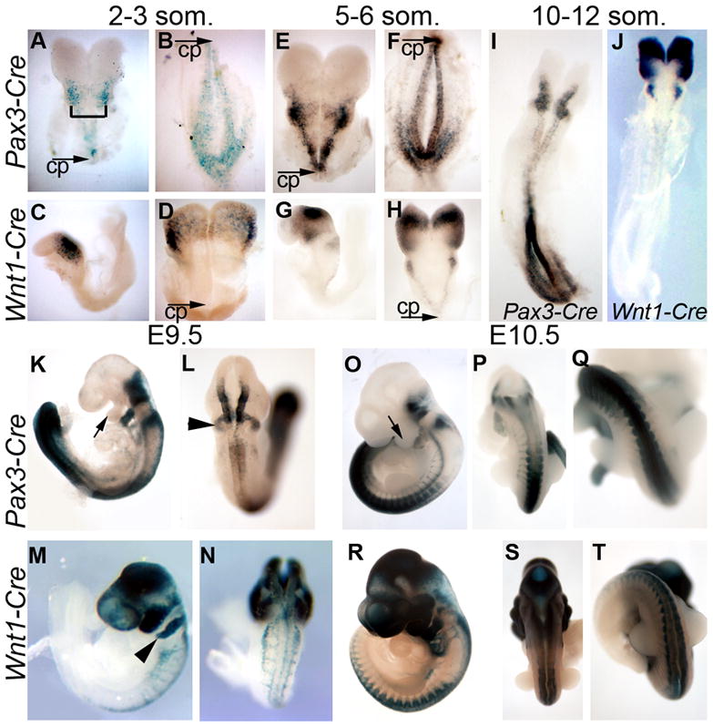
Figure 1. Spatiotemporal recombination patterns of Pax3-Cre and Wnt1-Cre.

Embryos carrying the R26R Cre reporter and either Pax3-Cre (A,B,E,F,I,K,L,O–Q) or Wnt1-Cre (C,D,G,H,J,M,N,R–T) were stained for β-galactosidase activity to mark cells which have undergone Cre-mediated recombination. Embryos at 2–3 somites (A–D) and 5–6 somites (E–H) were bisected at the initial closure point (CP) to allow visualization of the entire dorsal neural epithelium (bracket in A indicates hindbrain). At E9.5 (K–N) and E10.5 (O–T), embryos are shown in lateral (K, M,O,R) as well as dorsal views at the level of the thorax (L,N,P,S) or posterior to the forelimb bud (Q,T). Arrows (K,O) highlight the first pharyngeal arch, arrowheads (L,M) indicate the second pharyngeal arch.
