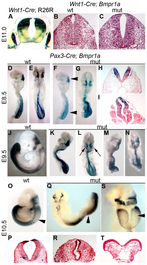
Figure 2. Neural tube phenotypes in Wnt1-Cre;Bmpr1a and Pax3-Cre;Bmpr1a mutants.
(A) Transverse section of E11.0 Wnt1-Cre;R26R embryo stained for β-galactosidase activity. Transverse sections of control (B) and Wnt1-Cre;Bmpr1a (C) neural tubes were stained with haematoxylin and eosin and show no neurulation defects in the Wnt1-Cre;Bmpr1a mutants. (D–T) Pax3-Cre ablation of Bmpr1a. (D–I; E8.5) Pax3-Cre; R26R control embryos (D lateral view, E dorsal view) have initiated neurulation in the spinal cord. Pax3-Cre;Bmpr1a;R26R embryos also initiate spinal neurulation but the neural tube is dysmorphic (F lateral, G, dorsal: arrowheads in F indicate regions shown in section in H,I). (J–N; E9.5) Pax3-Cre;R26R control embryo (J) has completed neurulation, whereas Pax3-Cre; Bmpr1a; R26R littermates fail to turn and often show hindbrain exencephaly (K,M lateral; L,N dorsal; arrows highlight hindbrain exencephaly). (O–T; E10.5) Pax3-Cre; R26R control embryo (O); Pax3-Cre; Bmpr1a; R26R mutant embryos show a failure to turn and mid-hindbrain exencephaly (Q,S). Arrowheads in O, Q, S indicate regions shown in section in P,R,T respectively.

