
Figure 1.
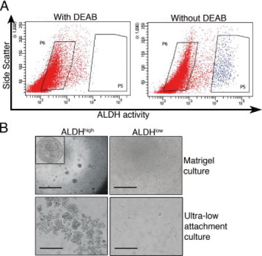
Isolation of primary renal cortex cells based on ALDH activity. A: Representative FACS analysis of a single-cell suspension of normal renal cortical tissue incubated with fluorescent ALDH substrate. The left panel shows the cellular fluorescence in the presence of the ALDH inhibitor diethylaminobenzaldehyde (DEAB), and the right panel shows the uninhibited reaction with the gate defining the ALDHhigh cells. The FACS gatings used are marked by boxes. In all the experiments, cells were first viability sorted by incubation in 7-amino-actinomycin D. B: The ALDHhigh population was enriched for cells with colony-forming capacity as assessed by plating in Matrigel culture (top row; scale bar = 1000 μm). Inset: A cellular sphere formed in the ALDHhigh culture at higher magnification (×40). The ALDHhigh population was also enriched for cells with the capacity to grow under ultra-low attachment conditions (bottom row; scale bar = 200 μm). The ALDHlow cells displayed none of these capacities.