
Figure 1.
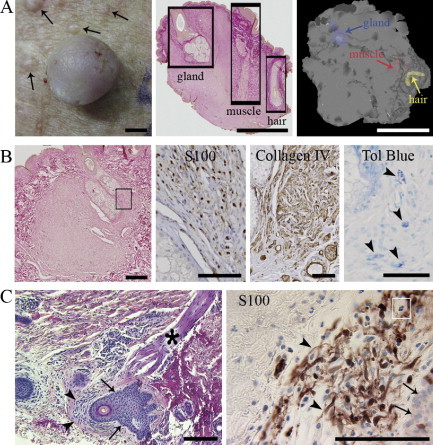
Association of hair follicles with neurofibromas at different stages. A: Cutaneous neurofibroma (diameter ∼7 mm) and numerous small, yet visible, tumor growths (left, arrows); all neurofibromas analyzed contained elements of a hair follicle (middle); three-dimensional model of a neurofibroma (right). Scale bars = 2 mm. B: Microscopic neurofibroma ensheathing a hair root (left). The rectangle depicts the approximate area further immunolabled for S100 and collagen IV; mast cells (arrowheads) are visualized by toluidine blue staining (right). Scale bars: 250 μm (left); 100 μm (S100, Collagen IV, and Tol Blue). C: Minute S100-positive neurofibroma intimately associated with hair follicle in apparently healthy looking skin of a patient with NF1. Tumor mass (arrowheads), follicular epithelium (arrows), erector pili muscle (asterisk), and mast cell (white rectangle) are shown. Scale bars = 200 μm.