
Supplemental Figure S1.
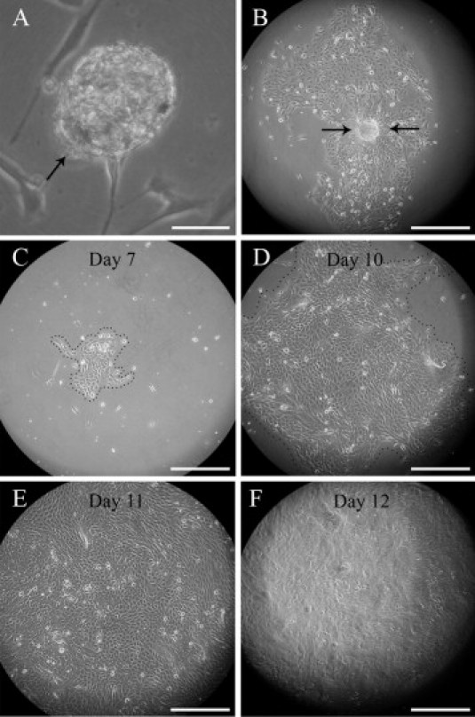
Proliferation of neurofibroma-derived cells in stem cell medium. A: A sphere of NFPs (arrow) anchored to the bottom of the culture disk. B: A single sphere has initiated the fast spreading cell sheet. C–F: A 7- to 12-day time series illustrates the fast proliferation rate of progenitor cells in a growing cell sheet. At day 7 (C), the cells are still confined to a limited area, but by day 10 they fill almost the entire optical field (D), clearly exceeding it by day 11 (E). On day 12 (F), the culture displays a marked difference of appearance as it becomes multilayered. See also Supplemental Videos S1 and S2. Scale bars: 50 μm (A); 300 μm (others).