
Figure 4.
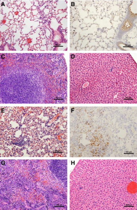
Anti-LcrV antibody treatment slows the progression of disease in Cxcr2−/− mice. Wild-type (WT) and Cxcr2−/− mice, treated with 400 μg anti-LcrV polyclonal antibody IP 60 minutes before intranasal challenge with 6000 CFU Y. pestis CO92 were euthanized 60 hours postinfection, and tissues were harvested and fixed in 10% formalin for histochemical and immunohistochemical analysis. WT lungs (A and B), spleen (C), and liver (D); Cxcr2−/− lungs (E and F), spleen (G), and liver (H). Immunohistochemistry with anti-Gr-1 was used to identify neutrophils (B and F). Images are shown at 20× magnification.