
Figure 3.
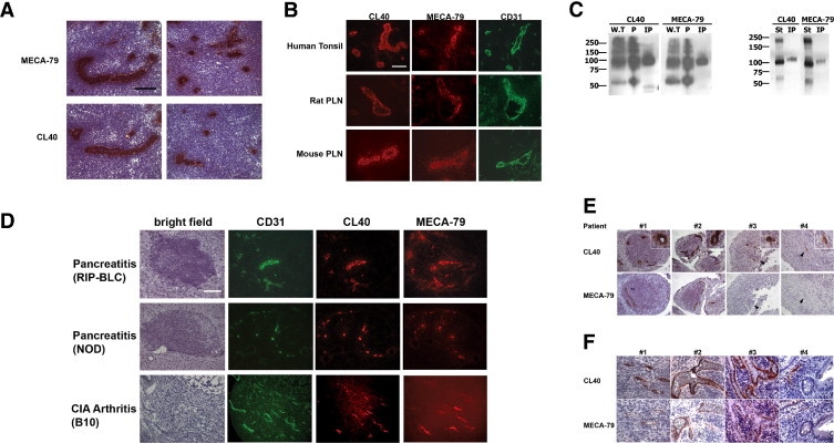
CL40 reactivity with high endothelial venules (HEVs) and HEV-like vessels and with PNAd components. A: Murine peripheral lymph node (PLN) sections were stained with MECA-79 and CL40 using horseradish peroxidase immunohistochemistry. B: Adjacent sections of human tonsil, rat PLN, and murine PLN were stained by with CL40 (red), MECA-79 (red), and a CD31 antibody (green, to reveal vessels) using fluorescence immunohistochemistry. C: Left: Human whole tonsil (W.T), peripheral lymph node addressin or PNAd (designated P) and immunoprecipitated human CD34 (IP) were electrophoresed. Right: Stroma from mouse PLN (St) and murine CD34 isolated by immunoprecipitation from a detergent lysate of stroma (IP) with anti-mouse CD34 was electrophoresed. The transferred membranes were blotted with MECA-79 or CL40 at 5 μg/ml. Data are representative of three independent experiments. D: Sections of pancreata from RIP-BLC and NOD mice, and the synovium from mice with collagen-induced arthritis (B10 mice) were sectioned. Consecutive sections were stained with hematoxylin (bright field), CL40 (red), MECA-79 (red), and a CD31 antibody (green). E: Adjacent sections of paraffin-embedded synovium from four patients with rheumatoid arthritis were stained with CL40 and MECA-79. Patient 4 exhibited CL40+ vessels that were not stained with MECA-79. In other patients, in addition to double-positive vessels, individual CL40+MECA-79− vessels are evident. Insets: Higher magnification views of CL40+ vessels. Arrowheads indicate corresponding vessels in adjacent sections of individual patients. F: Adjacent sections of paraffin-embedded colonic mucosa from patients with ulcerative colitis were stained with CL40 and MECA-79. All scale bars = 100 μm. Original magnification, 20×.