
Figure 6.
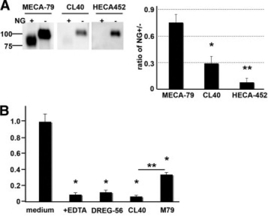
Contribution of N-glycans to CL40 reactivity. A: CD34 was immunoprecipitated from human tonsillar PNAd, treated with N-glycosidase F (NG) or PBS at 37°C for 42 hours, and then analyzed by Western blotting with the indicated antibodies. The band intensities were calculated as the ratios of NG-treated over untreated (right panel) using Multi Gauge software as described under Materials and Methods. The indicated means and SEM values were derived from three independent experiments. *P < 0.01 and **P < 0.005 against MECA-79, as determined by one-way analysis of variance and Tukey's post hoc test. B: Attachment of 300.19L cells to HEVs in cryosections of human tonsil was determined in the presence of 100 μg/ml of the indicated antibodies or medium alone. Isotype controls were also used in these experiments, although data are not shown because the results were the same as with medium only. As determined by one-way analysis of variance and Tukey's post hoc test, *P < 0.005 for the difference between the indicated treatment and medium alone and **P < 0.01 for the difference between CL40 and MECA-79. There was a significant difference between MECA-79 and EDTA at P < 0.01, and between MECA-79 and DREG-56 at P < 0.01.