
Figure 3.
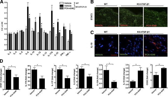
Effect of PCA-4248 on mRNA and/or protein levels of Th1-, Th2-, and Th17-related cytokines and transcription factor STAT3 and COX-2 in K5.hTGF-β1 transgenic mice. The WT or K5.hTGF-β1 transgenic mice were injected i.p. twice weekly for 4 weeks with PCA-4248 (10 mg/kg) or its vehicle. A: Serum samples were collected at the end of the 4-week treatment regimen from either vehicle-treated WT or vehicle- or PCA-4248–treated K5.hTGF-β1 transgenic mice. A multicytokine ELISA was performed to analyze the relative levels of IL-2, IL-4, IL-5, IL-6, IL-10, IL-12, IL-13, IL-17A, IL-23, interferon-γ, and TNF-α. Data shown are from one representative experiment, with n = 3 to 4 per treatment group. *P ≤ 0.05; **P ≤ 0.01, vehicle-treated K5.hTGF-β1 vs vehicle-treated WT mice. #P ≤ 0.05, PCA-4248–treated versus vehicle-treated K5.hTGF-β1 mice. O.D., optical density. Immunofluorescent staining of STAT3 (B) and IL-10 (C) (blue: DAPI; red: IL-10) protein in dorsal skin. Scale bars: 200 μm (B); 100 μm (C). D: Quantitative RT-PCR was performed at the end of the 4-week treatment regimen from either vehicle-treated WT or vehicle- or PCA-4248–treated K5.hTGF-β1 transgenic mice with RNA isolated from individual mouse dorsal skin for transcription factors STAT3 and transcripts encoding IL-17A, IL-17F, IL-6, IL-12A, IL-10, and COX-2. Data shown are from one representative experiment, with n = 5 to 7 per treatment group. *P ≤ 0.05. Error bars represent SEM.