
Figure 1. Xolloid (XLD) Ventralizes Ectoderm and Mesoderm.
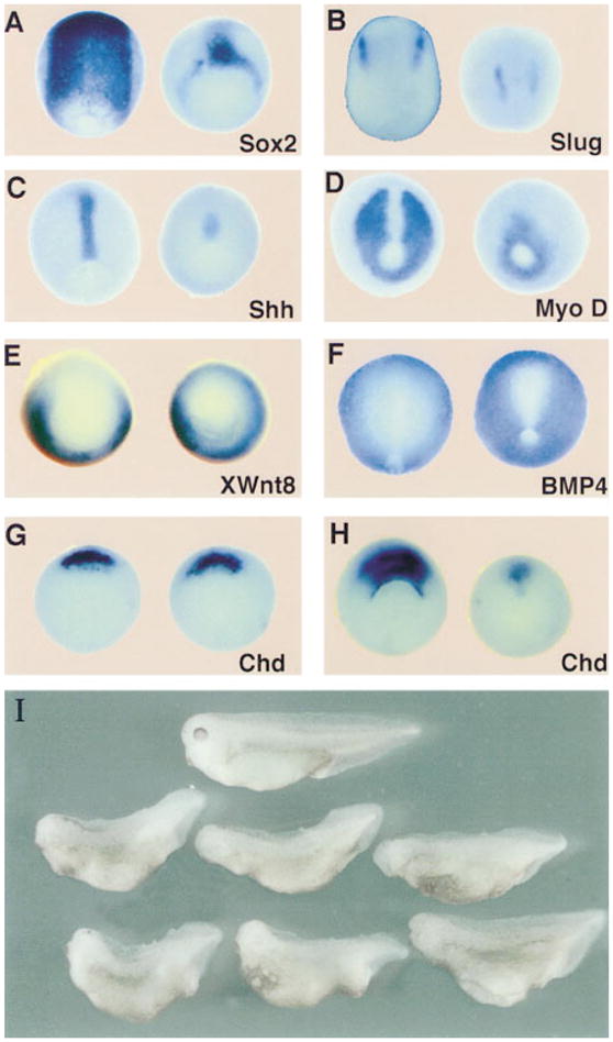
(A–H) Ventralization of ectoderm and mesoderm revealed by whole-mount in situ hybridization. Uninjected (left) and XLD mRNA–injected embryos (right) were probed for the indicated markers. Injections were done radially with 200 ng of XLD mRNA per blastomere at the 8-cell stage in the animal cap (A and B) or in the marginal zone (C–H). (A–D) Neurula stage embryos stained for (A) the neural marker Sox-2, (B) the neural crest marker slug, (C) the dorsal mesoderm and floor plate marker Shh, and (D) the dorsal mesoderm marker MyoD. Note the reduction of the neural and dorsal mesodermal fields. (E) At midgastrula stages, the ventrolateral marker Xwnt8 is expanded and expressed in the organizer in Xld-injected embryos. (F) At neurula stages, the BMP-4 domain is expanded in Xld-injected embryos, reducing the size of the neural plate. (G and H) Chd at early and late gastrula stages, respectively. Note that the transcription of Chd in Xld-injected embryos starts normally but collapses during the course of gastrulation.
(I) Normal (top) and XLD-injected 3-day tadpoles (bottom). Embryos were injected radially with 200 pg of XLD mRNA in each blastomere at the 4-cell stage. Note the recovery to a relatively mild ventralization (93% affected, DAI = 3.2, n = 311) when compared with the severe reduction in axial and neural markers observed at the early stages.