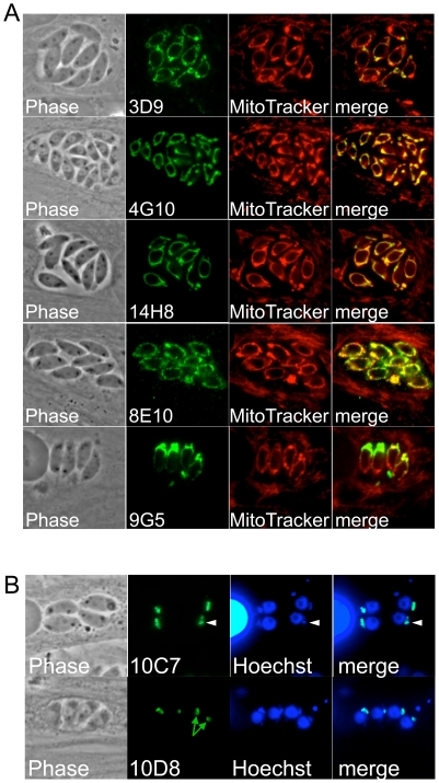
Figure 3. Antibodies detecting the parasite mitochondrion and apicoplast.
A) Antibodies detecting the parasite mitochondrion as assessed by Mitotracker colocalization which labels both the host and parasite mitochondria. Each of the monoclonals only stains the single tubular parasite mitochondrion and does not cross-react with the host organelle. B) 10D8 and 10C7 stain the apicoplast as detected by Hoechst co-staining. The apicoplast DNA is seen a single spot that is just anterior to the parasite nucleus (arrowheads). 10D8 shows an example of the central hole that lacks staining corresponding to the matrix of the apicoplast (green arrows).

