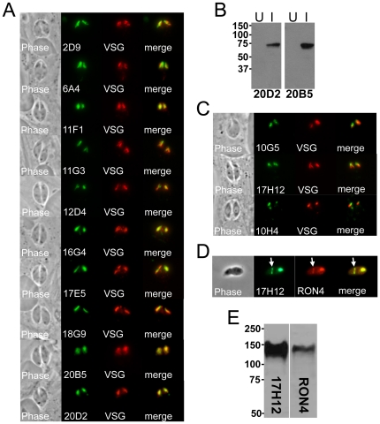
Figure 5. Antibodies against the rhoptry bodies and rhoptry necks.
A) Phase contrast and IFA analysis of rhoptry mAbs staining the body portion of the rhoptries. Rhoptry colocalization is shown using a Toxoplasma ROP1-VSG construct expressed in Neospora. B) mAbs 20D2 and 20B5 detect recombinant ROP4 expressed in E. coli. Western blot analysis of the identical strain uninduced (U) and induced (I) for ROP4 expression is shown. C) 17H12, 10G5, and 10H4 stain the more apical neck portion of the organelle and stain slightly apical to that of the ROP1-VSG fusion. A fourth RON protein detected by the mAb 8E3 was previously published [32] and is not shown here. D) 17H12 is secreted into the moving junction (arrow) in partially invaded parasites. The moving junction can be seen as a constriction of the parasite and by colocalization with cross-reactive sera against Toxoplasma RON4. E) Western blot analysis showing similar migration for RON4 and 17H12. RON4 is again detected by Toxoplasma cross-reactive antibodies against Neospora lysates.

