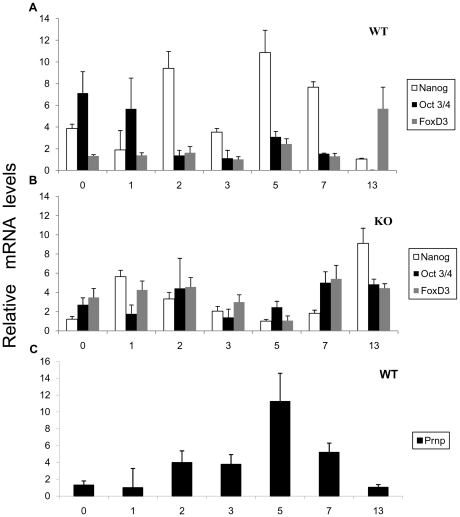
Figure 2. Pluripotency in the WT and Prnp-null cell lines.
WT EB mRNA (A) and KO EB mRNA (B) expression patterns of three pluripotency genes during differentiation. Note the lack in the KO line (B) of an increase in Nanog expression on Day 5. C) WT EB mRNA expression pattern of Prnp during differentiation (Days 0, 1, 2, 3, 5, 7 and 13). Prnp is expressed at its highest levels on Day 5 (Error bars, s.e.m.).

