
Fig. 4. Entry of P85 in primary rat cortical neurons visualized by confocal microscopy.
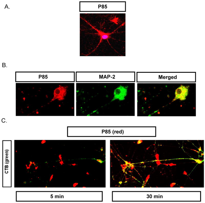
(A) Cells exposed to TRITC-P85 (red, 0.001% w/v) for 1 hr and then stained by DRAQ-5 (20 nM, 30 min.) to visualize nucleus. (B) Cells exposed to TRITC-P85 as in panel (A) and then fixed and stained by anti-MAP-2 antibody (10 μg/ml, 4° C overnight) and Alexa 488- labeled secondary antibody. (C) Cells exposed to TRITC-P85 (0.001% w/v) in the presence of Alexa 488-labeled CTB (5 μg/ml) for 60 min, washed and subjected to time lapse live cell imaging after 5 and 30 min.