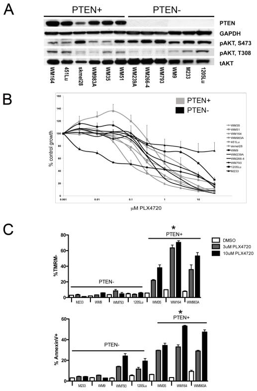
Figure 1. PTEN predicts for PLX4720-induced apoptosis.
(A) Basal PTEN and phospho-AKT (pAKT) (S473, T308) expression in PTEN+ (WM164, 451Lu, SK-mel-28, WM983A, WM35, WM51) and PTEN− (WM239A, WM266-4, WM793, M233, WM9, 1205Lu) melanoma cell lines. (B) MTT assay of PTEN+ (gray) expressing versus PTEN− (black) cell lines. (C) PTEN+ cells are more sensitive than PTEN− cells to PLX4720 mediated apoptosis. Cells treated for 48hrs with 3 or 10μM PLX4720 before being stained for TMRM and Annexin-V. Apoptosis was measured by flow cytometry. Data shows mean, +/− S.E. mean of three independent experiments. * Indicates PTEN+ cohort significantly different from PTEN− cohort (P<0.05).

