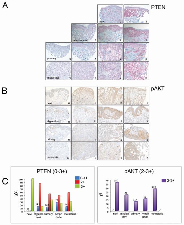
Figure 2. PTEN expression is lost at all stages of melanoma progression.
(A) Images showing representative immunohistochemical staining of PTEN and (B) pAKT expression in a tissue array of nevi, atypical nevi, primary and metastatic melanoma patient tumor samples. 0-1 indicates no to low PTEN expression and +3 indicates the highest expression while +2-3 relates to high expression of pAKT. Magnification x 200. (C) Left panel shows percentage loss of PTEN expression in each subset of patient samples as indicated in blue while the right panel shows AKT activity in matched samples.

