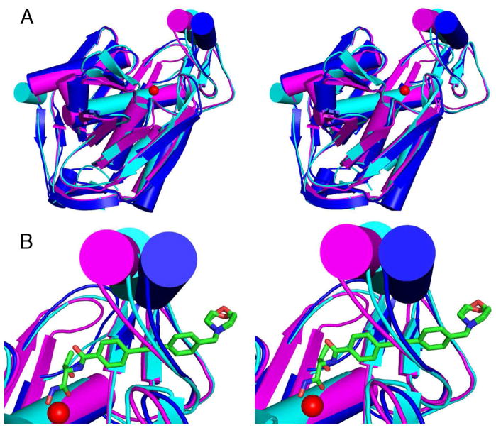
Figure 5.
(A) Superposition of YeLpxC (cyan), AaLpxC (magenta), and PaLpxC (blue). (B) Close-up view of the βαβ domain with the catalytic zinc ion shown as a red sphere and the CHIR-090 inhibitor shown as a green stick figure. This view highlights the apparent left-to-right movement of the βαβ domain, as well as the flexibility in-and-out of the plane of the paper.

