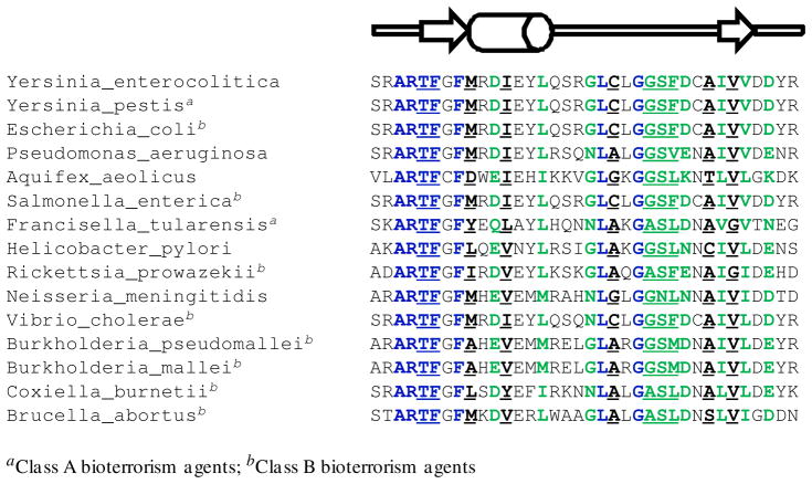
Figure 6.
Partial sequence alignment of LpxC (residues 187–222, YeLpxC numbering) from selected Gram-negative bacteria. Fully conserved residues are blue and largely conserved residues are green. The residues that interact with the CHIR-090 inhibitor through van der Waals or hydrogen bond interactions are underlined. A cartoon of the secondary structure highlights the βαβ domain (arrows represent the β-strands and the cylinder represents the short α-helix).

