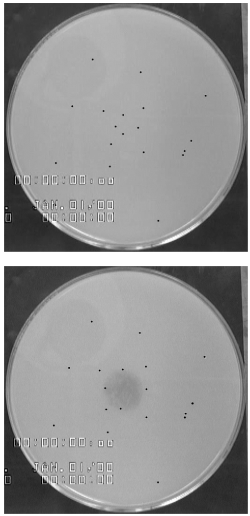
Figure 1.
Before and after images of a typical spreading experiment with a 2 µl droplet of a 2 mM SDS solution containing erythrosine B dye placed on the center of the PGM subphase. Each particle is marked by a black dot in the image for ease of viewing. For scale reference: the Petri dish is 14 cm in diameter. Note that innermost tracer particles are swept outward by the spread of the dye solution.

