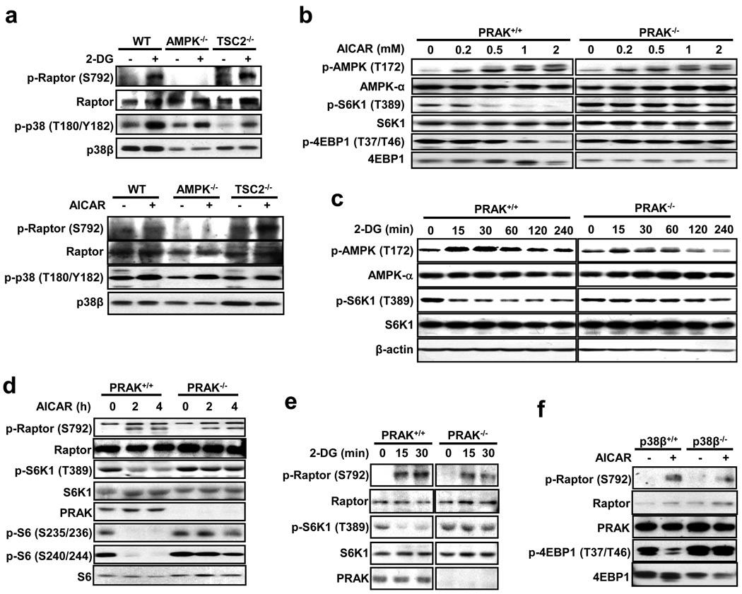
Figure 4.
PRAK's function in suppressing mTORC1 is independent from AMPK. (a) MEF cells with the indicated gene deletions were infected with a lentivirus expressing p38β and stimulated with or without 25 mM 2-DG for 30 minutes or 2 mM AICAR for 4 hours. Cell lysates were analyzed by immunoblotting for the protein and phosphorylation levels of raptor and p38β. (b) AICAR-induced AMPK phosphorylation is comparable in PRAK+/+ and PRAK−/− cells. PRAK+/+ and PRAK−/− MEF cells were stimulated with different doses of AICAR for 2 hours. Phosphorylation and protein levels of AMPK, S6K1, and 4EBP1 were determined by immunoblotting with corresponding antibodies. (c) 2-DG-induced AMPK activation in MEF cells is not affected by PRAK knockout. PRAK+/+ and PRAK−/− MEF cells were treated with 25 mM 2-DG for 0, 15, 30, 60, 120, or 240 minutes. Cell lysates were analyzed by immunoblotting for the levels of the indicated proteins and their phosphorylation states. (d) PRAK+/+ and PRAK−/− MEF cells were treated with 0.5 mM AICAR for 0, 2, and 4 hours. Cell lysates were analyzed by immunoblotting for the levels of the indicated proteins and their phosphorylation states. (e) 2-DG-induced raptor phosphorylation is not compromised in PRAK-deficient MEF cells. PRAK+/+ and PRAK−/− MEF cells were treated with 25 mM 2-DG for 0, 15, or 30 minutes. Cell lysates were analyzed by immunoblotting for the levels of the indicated proteins and their phosphorylation states. (f) AICAR-induced raptor phosphorylation is not compromised in p38β-deficient MEF cells. p38β+/+ and p38β−/− MEF cells were stimulated with 0.5 mM AICAR for 4 hours. Cell lysates were analyzed by immunoblotting for the levels of the indicated proteins and their phosphorylation states.

