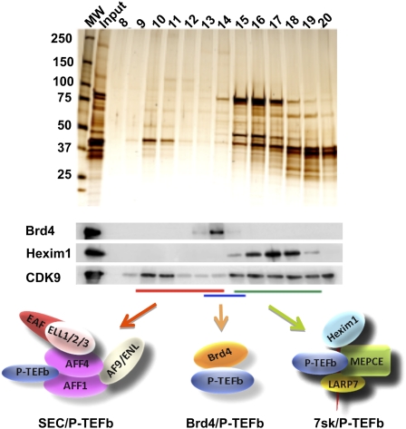
Figure 5.
Multiple P-TEFb complexes. P-TEFb, consisting of CDK9 and Cyclin T, is found in multiple complexes, which can be separated by gel filtration chromatography. CDK9-containing complexes from 293 cells were isolated by Flag affinity purification and subjected to size exclusion analysis. The resulting fractions were analyzed by silver staining and Western blotting. The inactive HEXIM1-containing P-TEFb complex, represented by HEXIM1, was enriched in fractions 15–19. The BRD4/P-TEFb complex peaked at fractions 13–15. SEC complexes are the largest P-TEFb complexes and are found in fractions 10–14, as described previously (Lin et al. 2010).

