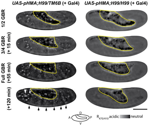
Fig. 5.
Cell-death-deficient embryos lack an acidified pHMA signal. Shown here are R470/410 ratio images of a UAS-pHMA;H99/TM6B (or UAS-pHMA;TM6B/TM6B) embryo (left column) and a UAS-pHMA;H99/H99 embryo (right column) injected with GAL4VP16 at ½, ¾, full and 65 minutes post GBR. Arrows indicate the normal pattern of segmental cell engulfment; the arrowhead points to an area with high macrophage density. Notice the lack of pHMA acidification in the H99 homozygous embryo. The yolk mass is outlined in yellow. Wide-field microscopy; 20X lens; lateral view; anterior, left; ventral, down; scale bar is 100 μm.

