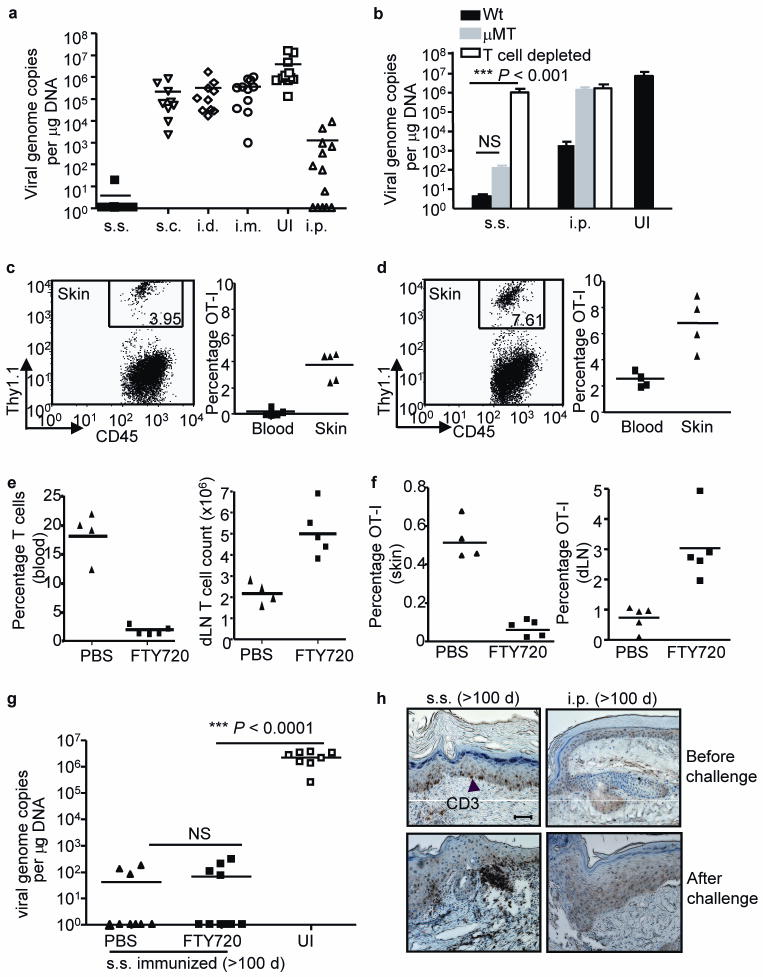
Figure 2. rVACV scarification generates superior protective immunity against cutaneous viral challenge that is mediated by skin-targeted TEM.
(a, b) Skin viral load measured by real-time PCR on d 6 after cutaneous challenge of Wt or μMT immune mice (immunized with VACV 6 weeks prior to challenge). In some Wt memory mice, T cells were depleted before and during the challenge (b). Data is pooled results from 3 independent experiments. (c, d) The frequencies of OT-I T cells in CD45+ leukocyte population in skin and blood either before (c) or after (d) challenge. FACS plots were gated on CD45+ leukocyte populations in skin. Numbers on the plots represent the frequencies of OT-I T cells in CD45+ populations. (e) T cell frequency in blood and absolute number in dLN measured immediately before secondary challenge to assess the efficiency of FTY720 blockage of T cell egress from lymphoid tissues. (f) Frequencies of OT-I cells in total viable cell populations in skin tissue and dLN at d 4 after challenge. (g) Cutaneous viral load at d 6 after secondary challenge determined by real-time PCR. Data are pooled results from 2 independent experiments. (h) Immunohistochemistry (IHC) staining showing the presence of CD3+ T cells in skin tissue of s.s. or i.p. immunized mice before or after secondary cutaneous rVACV challenge. Scale bar represents 5 μm. Photographs shown are the representative of 9 slides from 3 mice per group.

