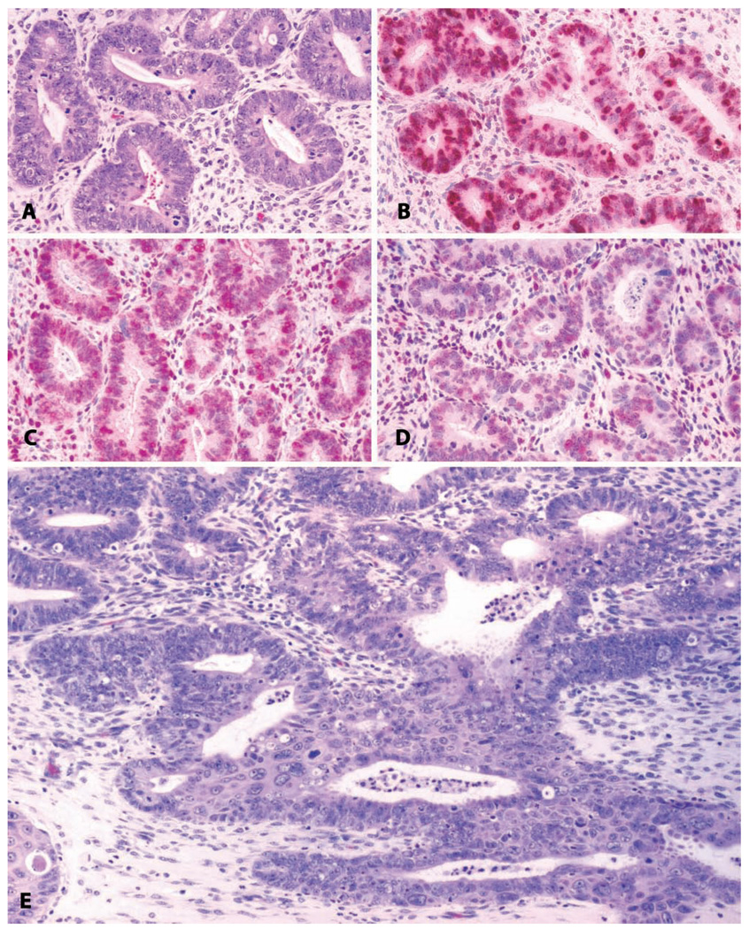
FIGURE 10.
Uterus: Hyperplasia with progression to invasive endometrial carcinoma. (A) Endometrial glandular hyperplasia in a thirty-two-year-old rhesus macaque chronically treated with estradiol by subcutaneous implant. “Complex hyperplasia” or “endometrial intraepithelial neoplasia.” H&E. (B) Corresponding area stained for proliferating cells; anti-Ki-67 antibody, Vector red chromogen, and hematoxylin counterstain. (C) Corresponding area stained for estrogen receptor alpha; anti-ER antibody, Vector red chromogen, and hematoxylin counterstain. (D) Corresponding area stained for progesterone receptors; anti-PR antibody, Vector red chromogen, and hematoxylin counterstain. (E) Adjacent region of the same slide; highly pleomorphic and invasive endometrial adenocarcinoma. H&E.

