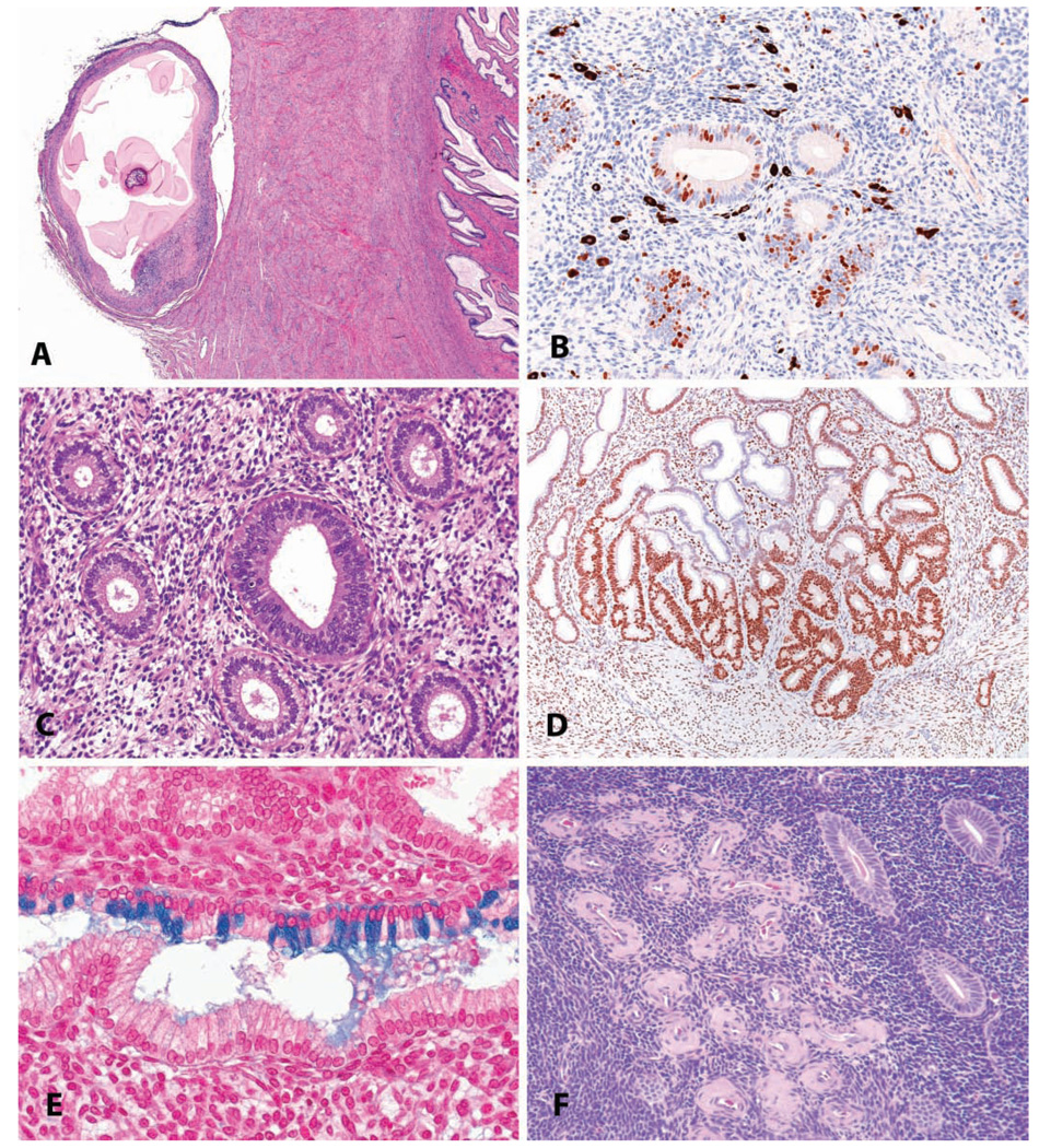
FIGURE 11.
Uterus: incidental findings. (A) Parasitic remnant manifest as a fibrous/mineralized cyst on the cervical serosa in a four-year-old cynomolgus monkey. H&E. (B) Endometrial melanosis in a four-year-old cynomolgus monkey, shown in an immunohistochemical stain for the proliferation marker. Immunohistochemistry: diaminobenzidine chromogen, hematoxylin counterstain. (C) “Luteal phase defect” in an adult cynomolgus monkey, with a mixture of follicular-phase features (round glandular lumens, pseudostratification) and luteal phase features (stro-mal proliferation and hypertrophy, glandular subnuclear vacuoles). (D) Focal glandular hyperplasia, basalis, in an adult cynomolgus monkey. Progesterone receptor immunostain, diaminobenzidine chromogen, hematoxylin counterstain. (E). Endometrial glandular mucous metaplasia in an adult cynomolgus monkey. Alcian blue stain. (F) Focal endometrial hyaline perivascular deposits in an aged cynomolgus monkey. H&E.

