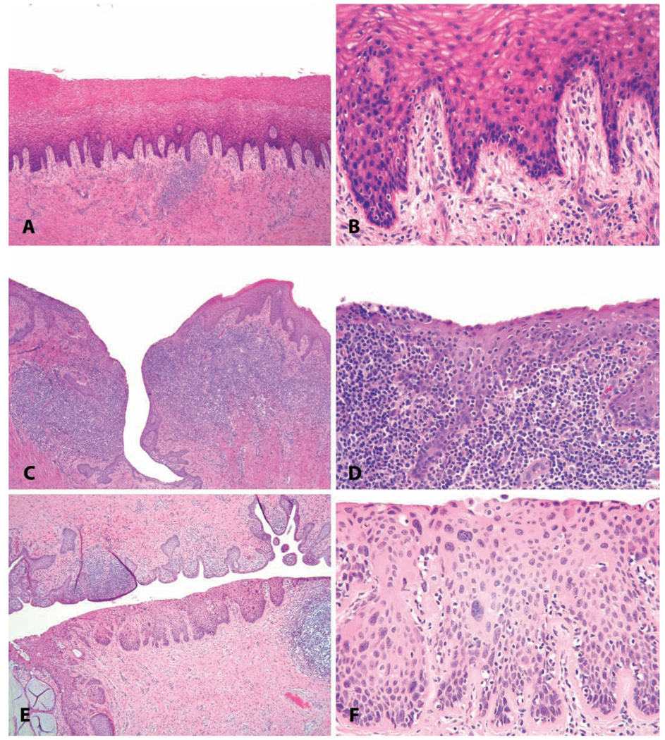
FIGURE 12.
Common vaginal lesions. (A–D) Vaginitis. (A and B) Minimal lymphocytic vaginitis, in a 22.5-year-old intact cycling animal with vaginal keratinization. (C and D) Severe chronic vaginitis with lymphoid follicular hyperplasia in a 7.5-year-old ovariectomized cynomolgus monkey (note atrophy of the surface epithelium and lack of keratinization). (E and F) Papillomavirus-induced in situ neoplasic lesion in a twenty-three-year-old cynomolgus monkey (cervical intraepithelial neoplasia, grade 2). H&E.

