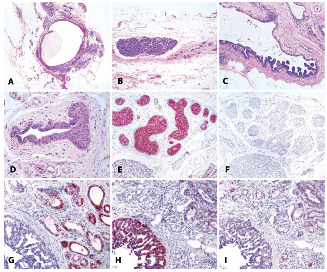
FIGURE 13.
Benign and malignant breast lesions. (A) Cystic change in a sixteen-year-old cynomolgus monkey, with marked dilatation of a large duct. H&E. (B) Focal lobular hyperplasia in the mammary gland of an adult cynomolgus monkey. Note the atrophy of other glandular elements on the right side of the photo. H&E. (C) Papillary ductal hyperplasia in an adult cynomolgus monkey. H&E. (D) Ductal hyperplasia with atypia in an eighteen-year-old cynomolgus monkey. H&E. (E and F) The same lesion as in (D) stained for estrogen receptor and the proliferation marker Ki67, respectively. Note overexpression of estrogen receptor. (G, H, and I) Ductal carcinoma in a five-year-old rhesus macaque, immunostained for (G) estrogen receptor, (H) proliferating cells/Ki67, and (I) progesterone receptor. Vector red chromogen; hematoxylin counterstain.

