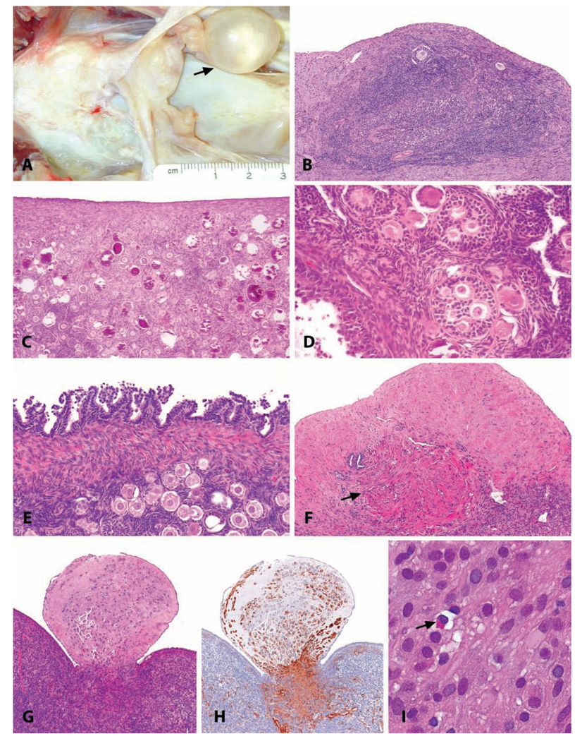
FIGURE 2.
(A) Gross appearance of a para-ovarian cyst in a nineteen-year-old cynomolgus monkey. (B) Ectopic ovarian tissue on the uterine serosal surface of an adult, normally cycling cynomolgus monkey. Note the presence of primary and atretic follicles in a dense ovarian-type stroma. H&E. (C) Multifocal mineralization in the ovary of a 2.5-year-old cynomolgus monkey. H&E. (D) Polyovular follicles in the ovary of a 2.5-year-old cynomolgus monkey. H&E. (E) Papillary hyperplasia of the ovarian surface epithelium in a 7.5-year-old cynomolgus monkey. H&E. (F) Focal smooth muscle metaplasia of the ovarian stroma in the ovary of an 18-year-old cynomolgus monkey; the absence of oocytes and fibrosis of the adjacent stroma reflects the age of the animal. H&E. (G, H, I) Ovarian deciduosis in a 16-year-old cynomolgus monkey, forming a polypoid mass projecting above the ovarian surface. (G) H&E. (H) anti-smooth muscle actin. (I) H&E at higher magnification. Typical endometrial lymphocytes were present (arrow).

