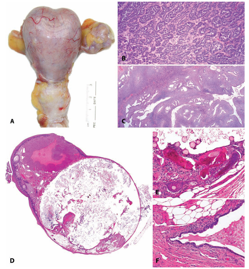
FIGURE 3.
Ovarian neoplasia. (A–C) Granulosa cell tumor from a twenty-six-year-old cynomolgus monkey with an estrogen-producing ovarian granulosa cell tumor, with the functional consequence of uterine enlargement by endometrial hyperplasia and myometrial hypertrophy. (A) Gross specimen. The uterus weighed more than 70 grams, compared to <2 grams for an ovariectomized animal or 7 grams for a cycling animal. (B) Typical histologic appearance of ovarian granulosa cell tumors. H&E. (C) Simple endometrial hyperplasia induced by estrogens of tumoral origin. H&E. (D–F) Benign ovarian teratoma in an 8.5-year-old cynomolgus monkey. (D) Subgross histologic appearance of this cystic neoplasm. H&E. (E, F) Multiple tissue types including hair, bone, fat, and glandular epithelium. H&E.

