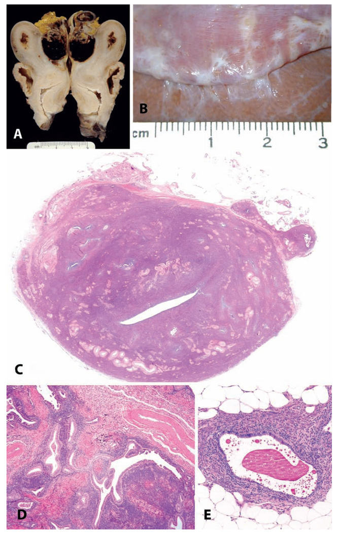
FIGURE 5.
Endometriosis and adenomyosis. (A and B) Variation in the gross appearance of endometriosis in macaques. (A) Typical blood-filled cyst caudal to the uterus in an adult female cynomolgus monkey. (B) Plaque-like pale lesions causing adhesions between the liver and diaphragm in a nineteen-year-old cynomolgus monkey. (C) Transverse section of the uterus of a twenty-four-year-old cynomolgus monkey with both adenomyosis and endometriosis, causing asymmetry of the uterus. H&E. (D and E) Typical histologic appearance of endometriosis, including endometrial glands, stroma, and hemorrhage or hemosiderin. H&E.

