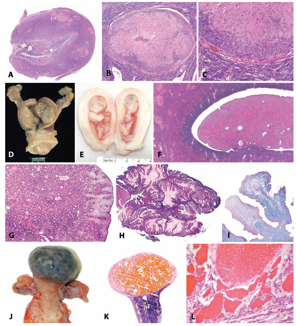
FIGURE 8.
Benign uterine neoplasms. (A) Longitudinal section through the uterus of a thirty-year-old rhesus macaque; within the myometrium there are four leiomyomas. H&E. (B and C) Higher magnification demonstrating smooth muscle morphology of the neoplastic cells. H&E. (D and E) Gross photographs of endometrial polyps in aged rhesus macaques; the polyps in (D) are sessile, and those in (E) are pedunculated. (F) Endometrial polyp consisting primarily of stromal tissue in a fourteen-year-old cynomolgus monkey. H&E. (G) Endometrial polyp in an adult rhesus monkey consisting primarily of glandular tissue. H&E. (H) Endometrial polyp in an adult rhesus monkey, with mucous differentiation. H&E. (I) Fibrosis within a polyp in an adult cynomolgus monkey; Masson’s trichrome stain. (J) Gross photograph of a myometrial cavernous hemangioma in a twenty-two-year-old cynomolgus monkey. (K) Subgross appearance. (L) Higher magnification. H&E.

