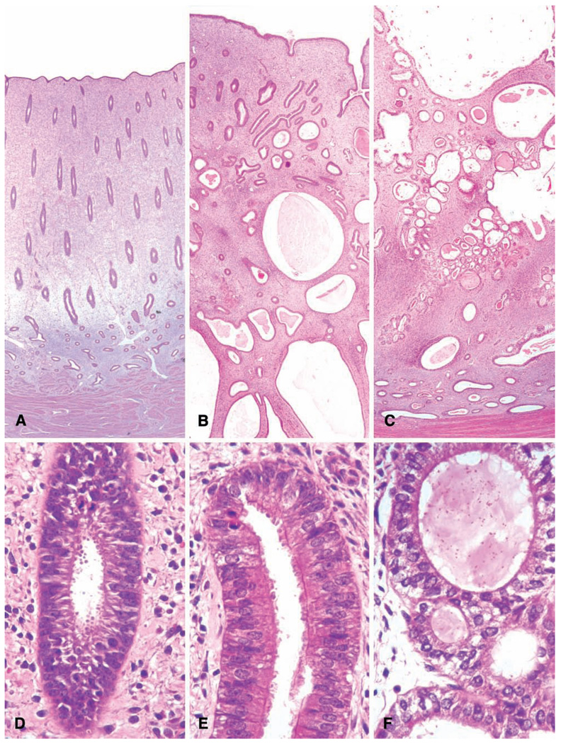
FIGURE 9.
Uterus: normal and pathologic proliferative changes. (A and D) Normal follicular phase endometrium with edema of the functionalis and orderly glandular proliferation. (B and E) Simple endometrial hyperplasia with disorganized glandular structures, cystic change, and ciliary metaplasia of the glandular epithelium. (C and F) Complex endometrial hyperplasia, with “back-to-back” crowding of endometrial glands.

