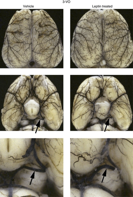
Figure 1.
Cerebral angioarchitecture. Visualization of the cerebral angioarchitecture using the latex method after maximal vasodilation. Dorsal view of brains investigated 1 week after three-vessel occlusion (3-VO) in vehicle- and leptin-treated animals. Leptin treatment leads to a moderate but not significant enlargement of ipsilateral PCA (arrows) compared with vehicle-treated animals.

