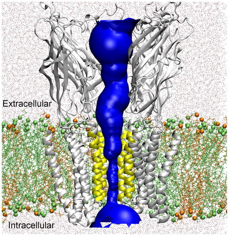
Figure 1.
Hybrid MD/continuum approach for calculating the single ion PMF through an equilibrated GLIC (PDB: 3EAM) protein. The channel pore for ion permeation is highlighted in blue. For the constricted TM pore (lined by the yellow TM2 segments), the single ion PMF was calculated via MD simulation. For other regions (i.e., the extracellular and intracellular domains), the single ion PMF was evaluated using a continuum dielectric approach. In the MD simulations, GLIC was embedded into a binary lipid containing POPE (lime) and POPG (orange) (in a ratio 3:1) and solvated by TIP3P water (dots). Lime spheres and orange spheres show the phosphorus atoms of POPE and POPG. For clarity, only four protein subunits are shown.

