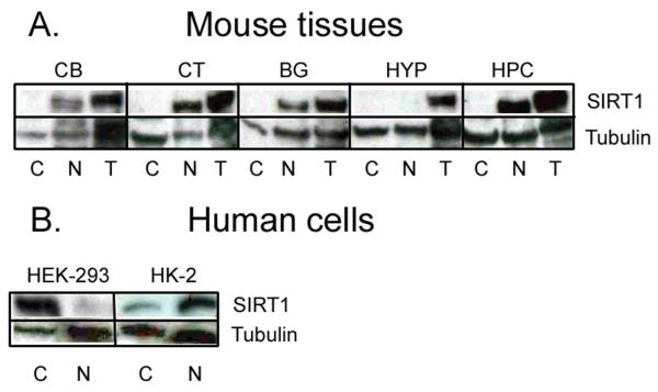
Fig. 3.
Representative Western blots of SIRT1 in rodent (A) CB = Cerebellum; CT = Prefrontal cortex; BG = Basal ganglia; HYP = Hypothalamus; HPC = Hippocampus. Note the relative absence of SIRT1 in mouse hypothalamus. Western blots of SIRT1 in human embryonic kidney cells (B). C = Cytoplasm; N = Nucleus; T = Total (i.e., both cytoplasmic and nuclear extracts). In human embryonic kidney (HEK-293) cells, SIRT1 is predominantly confined to the cytoplasm. This cytoplasmic expression, however, is reversed to the nucleus in adult human kidney (HK-2) cells. Tubulin was used to verify equal loading of proteins.

