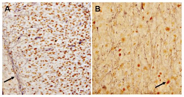
Fig. 8.
Representative bright-field photomicrographs depicting characteristic SIRT1-immunoreactivity in the cell nucleus and TH-labeling of neuronal fibers terminating within receptive DA fields of the rat fundus striate (A; Bregma 0.70 mm; Paxinos and Watson, 1986) and nucleus accumbens (B; Bregma 0.70 mm; Paxinos and Watson, 1986). Arrow in A points to neuronal fibers, whereas arrow in B points to cell nucleus labeled positive for SIRT1. A and B = 20X magnification.

