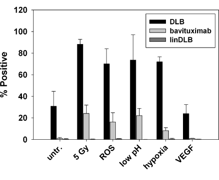
Figure 3.
Multiple stresses associated with the tumor microenvironment induce increased PE exposure on cultured EC. Flow cytometry was used to assay PE levels on ABAE cells after 1) treatment with 10 µM H2O2 to simulate ROS, 2) incubation in medium at pH 5.8 for 24 hours, 3) incubation under hypoxic conditions (1% O2, 94% N2, 5% CO2) followed by reoxygenation for 4 hours (5% CO2 in air), and 4) treatment with 20 ng/ml VEGF. Irradiation at 5 Gy was used as a positive control. Bavituximab was used to detect PS externalization. Rituxan gave no staining (not shown). LinDLB was used as a negative control for DLB.

