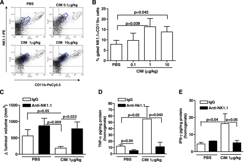
Figure 4.
Analysis of the role of NK cells on melanoma after CIM administration. CIM significantly increased the number of CD11b+CD11c+ NK1.1+ cells in melanoma tissue compared with PBS (n = 7). Representative plots are shown in A and mean data are depicted graphically in B. (C) Depletion of NK1.1+ cells (black bars) completely blocked the antitumor effect of CIM (1 µg/kg) (n = 7). Depletion of NK1.1 cells (black bars) attenuated CIM induction of TNF-α (D) and IFN-γ (E) levels in melanoma tissue homogenates (n = 7). All data are expressed as mean ± SEM. Statistical differences were determined by either two-tailed Student's t testor one-way ANOVA followed by Dunnett post hoc analysis as appropriate.

