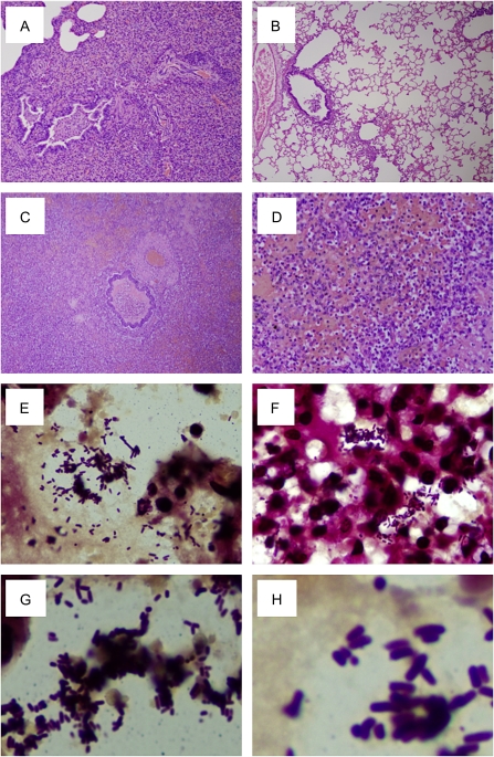
Figure 6.
Histopathology and immunohistochemistry of 2009 pandemic influenza A(H1N1) virus–infected ferret respiratory tissue. Representative photomicrographs of hematoxylin-and-eosin-stained tissue sections and Brown and Hopps tissue Gram-stained sections to detect bacteria from ferrets infected with wild-type or NAI resistant viruses at 14 days after inoculation or 12 days after exposure of the contact animals. A and B, Chronic changes observed in recovering animals included occasional examples of bronchiolitis obliterans with organizing pneumonia (A) and residual chronic active bronchiolitis and focal, mild alveolitis (B) (virus B inoculated ferret); original magnification 40×). One ferret found dead 12 days after exposure by contact transmission with virus B demonstrated pathology consistent with an acute bacterial pneumonia with destruction of the pulmonary architecture and a massive inflammatory infiltrate consisting predominantly of neutrophils (C, D) (original magnification 40× [A], 200× [B]). (E–H) Tissue Gram stain revealed abundant Gram-positive short rods, morphologically consistent with Listeria monocytogenes(original magnification 1000×).

