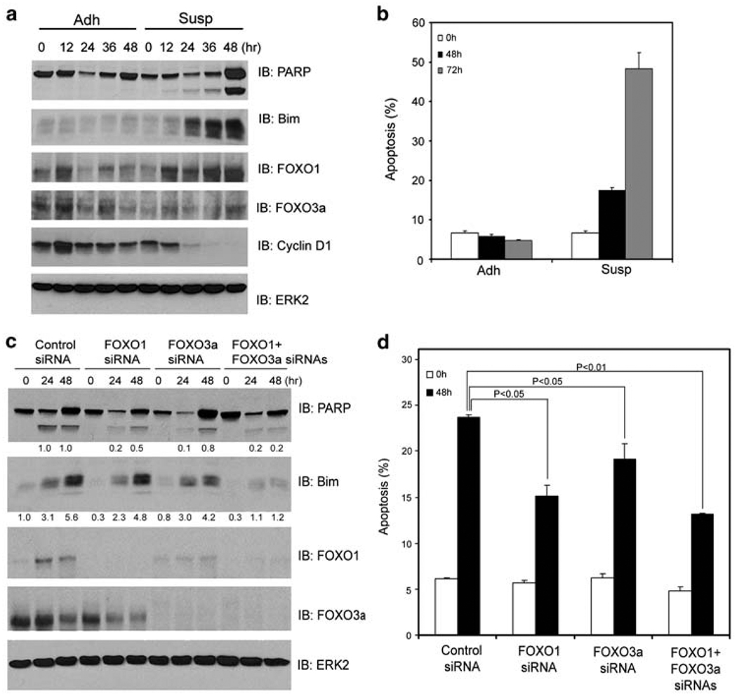
Figure 1.
FOXO1 and FOXO3a promote anoikis. DU145 cells grown in adhesion (Adh) or suspension (Susp) were collected at the indicated time points and subjected to immunoblotting analysis for expression of PARP, Bim, FOXO1, FOXO3a, and cyclin D1 proteins (a) and measurement of apoptosis (b). (c, d) DU145 cells were transfected with control siRNA, or siRNAs for FOXO1, FOXO3a, or both. At 48 h after transfection, cells were plated in adhesion or suspension and at the indicated time points cells were subjected to immunoblotting (c) and apoptosis (d) analysis. ERK2 was used as a loading control. Results from three independent experiments were quantified for apoptosis. Error bars indicate S.D. among three individual experiments

