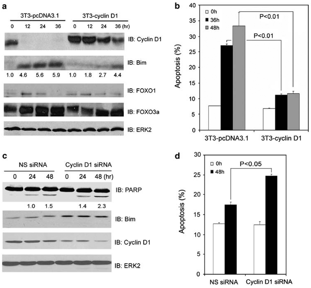
Figure 2.
Cyclin D1 inhibits anoikis. (a, b) NIH3T3 cells stably expressing either an empty vector (3T3-pcDNA3.1) or cyclin D1 (3T3-cyclin D1) were cultured in suspension and at the indicated time points cells were subjected to immunoblotting (a) and apoptosis (b) analysis. (c, d) LNCaP cells were transfected with a pool of cyclin D1-specific siRNAs or nonspecific control siRNAs. At 48 h after transfection, cells were cultured in suspension and at the indicated time points cells were subjected to immunoblotting (c) and apoptosis (d) analysis. The number underneath each band in the immunoblot indicates the relative intensity of the corresponding band. Error bars indicate S.D. among three individual experiments

