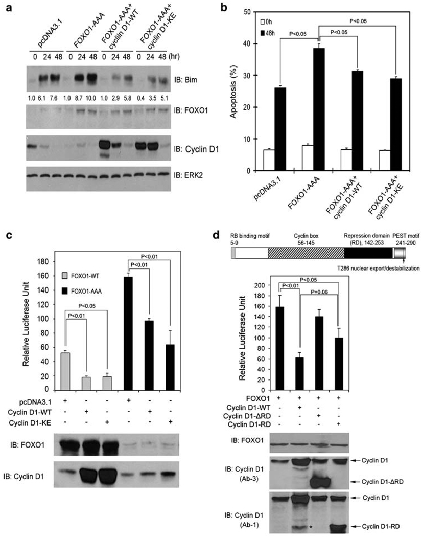
Figure 3.
Cyclin D1 inhibits FOXO1-augmented anoikis and FOXO1’s transcriptional activity. (a, b) DU145 cells were transfected with plasmids as indicated. At 36 h after transfection, cells were cultured in suspension and at the indicated time points cells were subjected to immunoblotting (a) and apoptosis (b) analysis. The number underneath each band in the immunoblot indicates the relative intensity of the corresponding band. (c) LNCaP cells were transfected with firefly and Renilla luciferase reporter constructs and plasmids as indicated. At 36 h after transfection, cells were subjected to luciferase activity measurement as described in ‘Materials and Methods’ (upper panel) or western blot analysis (lower panel). Error bars indicate S.D. among three individual experiments. (d) Top, a schematic diagram of the cyclin D1 protein shows its different functional domains. Middle and bottom, LNCaP cells were transfected with the indicated plasmids and luciferase activities and western blots were analyzed as described in (c). The asterisk indicates a nonspecific immunoreactive band

