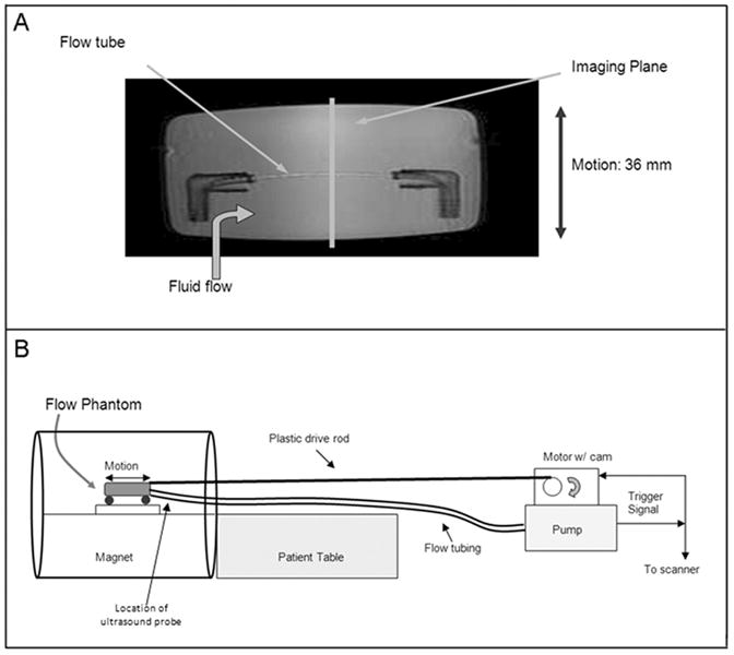
Figure 2.

(A) A 6mm diameter flow tube was embedded in gelatin in a plastic container which could be moved in a back-and-forth manner by a motor triggered in synchrony with the flow. This phantom provides a simulation of both motion and pulsatile flow as exhibited by the coronary arteries. (B) The flow pump and motor were placed two meters beyond the foot of the patient table with the plastic drive shaft and flow tubing running to the phantom in the magnet. The flow pump triggered the scanner and the motor for synchronized flow, motion, and acquisition.
