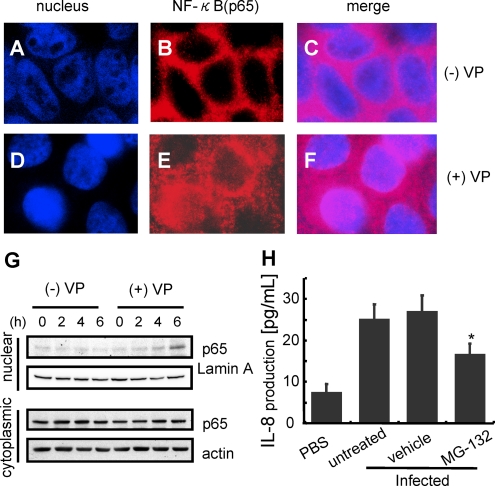
Figure 5.
V. parahaemolyticus induced activation and translocation of NF-κB. Caco-2 cells were uninfected (A - C) or infected with V. parahaemolyticus (D - F). The cells were fixed with 4% paraformaldehyde for 10 minutes and treated with .1% Triton X - 100 for 7 minutes. The fixed cells were incubated with anti-NF-κB antibody (1/500) in PBS containing 3% BSA. After treatment with the respective primary antibody, the cells were incubated with secondary antibody conjugated with Alexa fluor 568 (1/200) in PBS containing 3% BSA (B, E). Nuclei were stained with 500 nM DAPI followed by incubation with secondary antibody (A, D). G, Caco-2 cells were infected with V. parahaemolyticus (+VP) or uninfected (-VP) for the indicated times. H, After infection, nuclear extracts and cytoplasmic extracts were isolated from the infected cells, and the translocation level of NF-κB was shown by Western blotting. MG132 (25 μM), an NF-kB inhibitor, was added to the culture medium for 60 minutes prior to infection. IL-8 secretion into the medium was measured by enzyme-linked immunosorbent assay (ELISA). The results are shown as the mean ± SD calculated from 3 independent experiments. Statistical significance: *, P < .05 compared with untreated.

